X-Rays
An X-ray machine is a medical device used to produce images of the inside of the body using X-ray radiation. It is commonly used for diagnostic purposes to visualize bones, organs, and tissues, helping to identify injuries, diseases, or abnormalities.
Key points about X-rays include:
- Medical Imaging: X-ray machines are primarily used in medical settings to produce images of the internal structures of the human body. They are commonly used for diagnosing various conditions, such as fractures, lung infections, and dental problems.
- X-ray Generation: X-rays are a form of electromagnetic radiation that can pass through the body and produce an image on a film or digital sensor. X-ray machines generate X-rays by accelerating electrons through a high voltage and directing them towards a target material, typically tungsten.
Why X-rays are helpful?
- Medical Diagnostics: X-ray imaging is widely used in medical diagnostics to visualize the internal structures of the human body. It helps in detecting and diagnosing various conditions such as fractures, tumors, lung diseases, dental issues, and foreign objects swallowed or lodged in the body. X-rays can provide valuable information to physicians, enabling them to make accurate diagnoses and develop appropriate treatment plans.
- Non-Invasive: X-rays are a non-invasive imaging technique, meaning they can provide valuable information without the need for surgical procedures. This makes them a preferred method for diagnosing many medical conditions, as they eliminate the risks and complications associated with invasive procedures.
Preparation:
- Safety precautions: Before starting any preparation, make sure you are familiar with the safety guidelines and protocols associated with operating X-ray equipment. Wear appropriate personal protective equipment (PPE) such as lead aprons and gloves to minimize exposure to radiation.
- Check the machine: Inspect the X-ray machine to ensure it is in good working condition. Look for any visible damage or malfunctions. Verify that all cables, connectors, and components are securely attached.
Procedure:
- Patient Preparation: The patient is usually required to change into a hospital gown and remove any jewelry, accessories, or clothing items that could obstruct the X-ray image. The patient may also be asked to remove metal objects such as belts, coins, and keys.
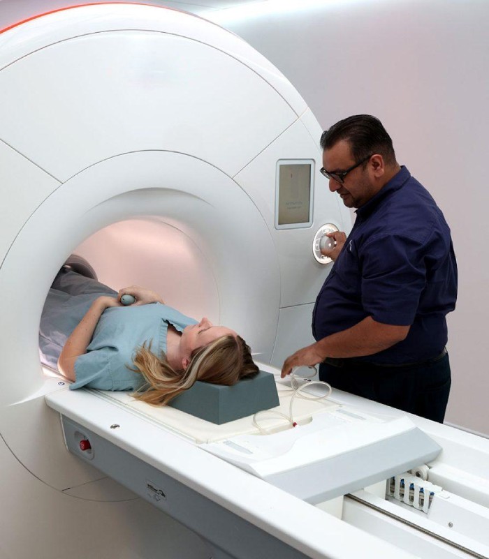
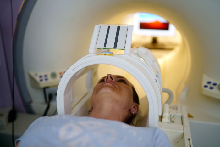
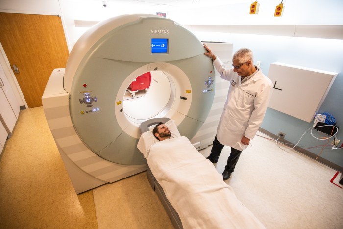
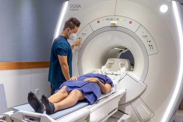
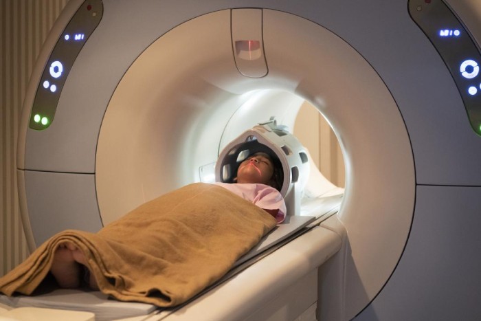
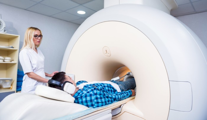
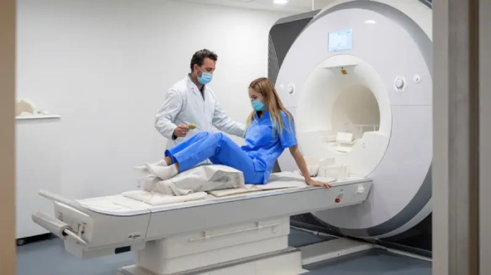
- Positioning: The patient is positioned by the radiologic technologist (X-ray technician) on a table or standing against a surface, depending on the type of X-ray being performed and the body part under examination. The technician may use pillows or other supports to help the patient maintain the necessary position.
Safety:
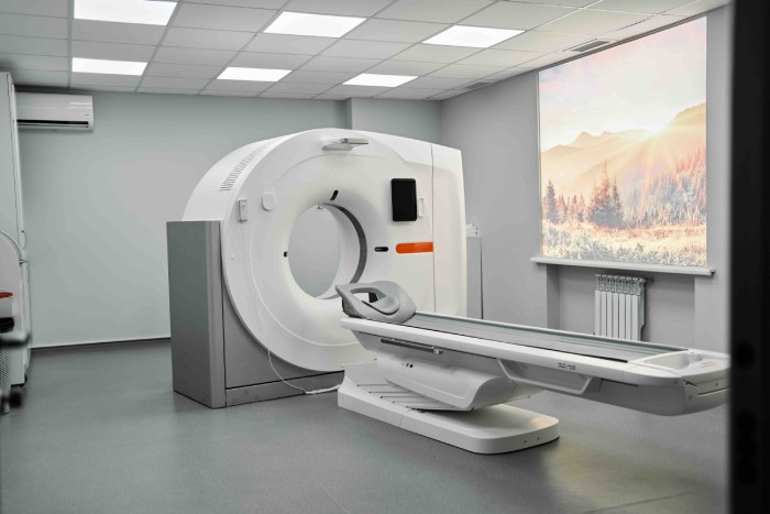
- Shielding: X-ray machines should be properly shielded to prevent radiation exposure to individuals in the vicinity. This includes lead-lined walls, floors, and ceilings in the X-ray room. Shielding is designed to absorb and block X-rays, ensuring that radiation is contained within the designated area.
- Restricted access: Access to the X-ray area should be limited to authorized personnel only. Clear signage and controlled entry should be implemented to prevent unauthorized individuals from entering the area during X-ray operations.
Diagnostic Applications:
- Detecting Fractures and Bone Abnormalities
- Evaluating Chest Conditions
- Dental Imaging
- Diagnosing Abdominal Conditions
- Assessing Joint Conditions
- Detecting Breast Abnormalities
- Identifying Gastrointestinal Disorders
- Locating Foreign Objects
It’s important to note that while X-rays are widely used for diagnostic purposes, they do involve exposure to ionizing radiation. Therefore, healthcare professionals take precautions to minimize radiation exposure to patients and themselves, ensuring that the benefits of the procedure outweigh the potential risks.
